
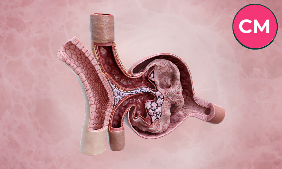

If you’ve ever stared at a histology slide wondering which pink blob is supposed to be the “filtration unit” everyone keeps talking about, you’re not alone. The renal corpuscle is one of those structures that sounds complicated in a textbook but makes total sense once you see it broken into its actual parts. In simple terms, the renal corpuscle is the tiny, ball-shaped filtering unit inside each nephron of the kidney, and it’s where the entire process of urine formation begins. This guide walks through its location, structure, function, and what happens when it stops working properly, without the dense jargon that makes most anatomy resources hard to get through.
What Is a Renal Corpuscle?
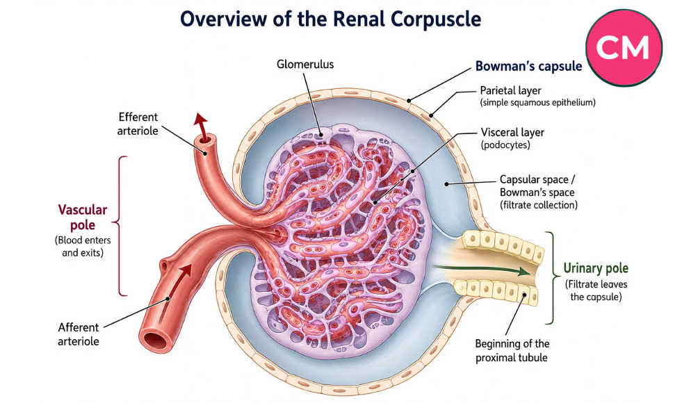
The renal corpuscle is the blood-filtering component of the nephron, the kidney’s functional unit. It’s made up of two main structures: the glomerulus (a tuft of capillaries) and Bowman’s capsule (a cup-shaped structure that surrounds it). Blood enters the glomerulus under pressure, and water, ions, glucose, and small waste molecules get pushed out of the capillaries into Bowman’s capsule, forming what’s called glomerular filtrate. This filtrate then travels into the renal tubule, where it’s gradually turned into urine.
What Is the Renal Corpuscle, Exactly?
Anatomically, the renal corpuscle sits at the very start of the nephron, the microscopic tubular unit that does the actual work of filtering blood. It’s also known by an older name, the Malpighian corpuscle or Malpighian body, a nod to the Italian anatomist who first described these structures centuries ago.
Each kidney contains roughly a million of these tiny corpuscles, though estimates in different textbooks range anywhere from one to four million depending on how the counting is done. That variation alone tells you something useful: kidney anatomy isn’t perfectly uniform from person to person, and even scientific sources don’t always agree on exact figures.
What makes the renal corpuscle interesting is that it’s essentially a pressure-driven sieve. Unlike other filtering systems in the body that rely on active transport, the renal corpuscle uses blood pressure itself to force fluid out of the capillaries. That’s part of why blood pressure control matters so much for kidney health, a connection that often gets glossed over in basic anatomy lessons.
Where Is the Renal Corpuscle Located?
The renal corpuscle is found exclusively in the renal cortex, the outer layer of the kidney. This is actually a handy identification trick if you’re ever looking at a kidney cross-section or a histology slide: if you see round, grainy-looking clusters, you’re looking at the cortex. The renal medulla, the inner portion, is made up of tubules and collecting ducts running in straight lines, so it looks striped rather than grainy.
Depending on where in the cortex a renal corpuscle sits, it belongs to either a cortical nephron (closer to the outer capsule, with a short tubule system) or a juxtamedullary nephron (closer to the medulla, with a much longer tubule that dips deep into the kidney). This distinction matters clinically because juxtamedullary nephrons play a bigger role in concentrating urine.
Renal Corpuscle Anatomy: The Parts That Make It Work
The renal corpuscle isn’t a single structure but a small system made of a few distinct parts working together.
The Glomerulus
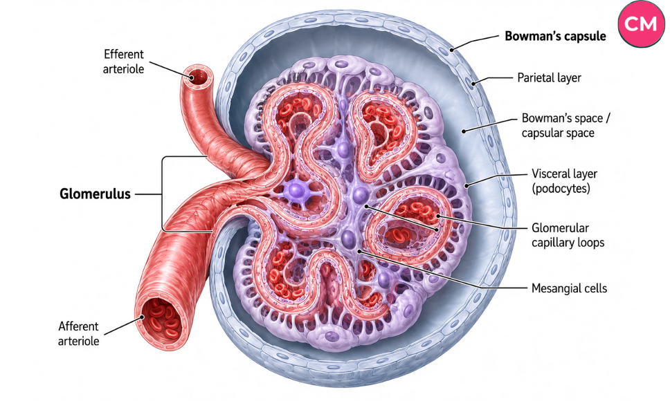
The glomerulus is a tangled network of capillaries that receives blood from a small vessel called the afferent arteriole. Because the afferent arteriole is wider than the efferent arteriole (the vessel that carries blood away), pressure inside the glomerulus stays high, which is exactly what’s needed to push fluid out of the blood and into the filtration system.
Bowman’s Capsule
Bowman’s capsule (also called the glomerular capsule) is the cup-shaped sac that wraps around the glomerulus. It has two layers:
- Parietal layer: the outer wall, made of simple squamous epithelium, mostly structural
- Visceral layer: the inner layer, made of specialized cells called podocytes that wrap around the capillaries and control what gets filtered
Between these two layers sits Bowman’s space, where the filtered fluid collects before heading into the renal tubule.
Mesangial Cells
Tucked between the capillary loops of the glomerulus, mesangial cells act like the support crew of the renal corpuscle. They help regulate blood flow through contraction, clear out trapped debris, and contribute structural support to the whole capillary tuft. They don’t get much attention in basic anatomy classes, but they’re essential to keeping the filtration system stable.
The Two Poles of the Renal Corpuscle
Every renal corpuscle has two functional “ends,” often described as poles:
| Pole | Location | What Happens There |
| Vascular pole | Where the afferent and efferent arterioles enter/exit | Blood enters and leaves the glomerulus here |
| Urinary (tubular) pole | Opposite side of the corpuscle | Filtrate exits into the proximal convoluted tubule |
This side-by-side comparison is something a lot of resources mention in passing without ever laying it out clearly, but understanding the two poles makes the whole filtration pathway much easier to visualize.
How the Renal Corpuscle Filters Blood: Step by Step
Here’s the actual sequence of events, broken down simply:
- Blood flows into the glomerulus through the afferent arteriole.
- High pressure inside the capillaries forces plasma (minus large proteins and blood cells) through the filtration barrier.
- The filtration barrier, made up of the capillary endothelium, the basement membrane, and podocyte filtration slits, blocks large molecules like albumin while letting water, ions, glucose, and small waste products through.
- This filtered fluid, now called glomerular filtrate, collects in Bowman’s space.
- Filtrate exits through the urinary pole into the proximal convoluted tubule, where reabsorption and further processing begin.
- Remaining blood (now minus the filtered fluid) exits through the efferent arteriole and continues to the peritubular capillaries.
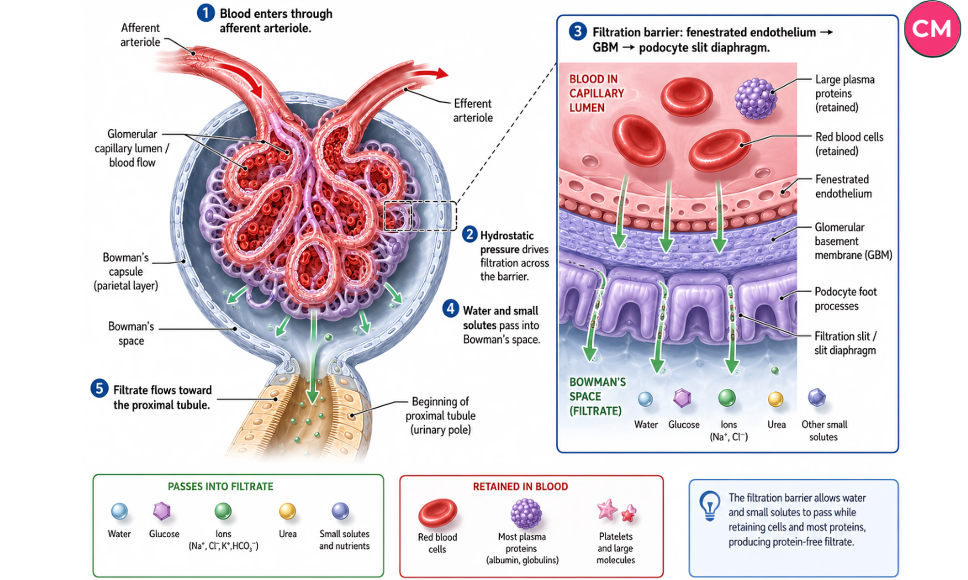
The filtration slits mentioned in step 3 are remarkably small, generally in the range of 20 to 30 nanometers wide, and the capillary pores themselves average around 70 nanometers. That’s a big part of why the filtration barrier does such a good job of holding onto essential proteins while letting waste pass through.
Renal Corpuscle vs Glomerulus vs Nephron vs Bowman’s Capsule
One of the most common points of confusion, especially for students first encountering kidney anatomy, is figuring out how these four terms relate to each other. Here’s a clear breakdown:
| Term | What It Actually Refers To |
| Nephron | The entire functional unit of the kidney, including the renal corpuscle plus the full tubule system |
| Renal corpuscle | The filtration portion of the nephron only, made of the glomerulus and Bowman’s capsule |
| Glomerulus | Just the tuft of capillaries inside the renal corpuscle |
| Bowman’s capsule | Just the cup-shaped capsule surrounding the glomerulus |
So the renal corpuscle is a component of the nephron, and the glomerulus and Bowman’s capsule are both components of the renal corpuscle. Getting this hierarchy straight early on saves a lot of confusion later, especially in physiology courses that reference these terms interchangeably.
What Happens When the Renal Corpuscle Is Damaged
When the renal corpuscle’s filtration barrier gets compromised, whether from disease, inflammation, or chronic conditions like diabetes or hypertension, the effects show up fairly quickly in urine and blood tests.
Some common consequences include:
- Proteinuria: protein (especially albumin) leaking into the urine because the filtration slits are no longer doing their job properly
- Hematuria: blood cells appearing in urine, often a sign of damage to the capillary walls
- Glomerulonephritis: inflammation of the glomerulus, which can be acute or chronic
- Nephrotic syndrome: a cluster of symptoms including heavy protein loss, swelling, and low blood protein levels, often linked to podocyte damage
Clinically, doctors often diagnose these conditions through a kidney biopsy, examining renal corpuscles under a microscope (sometimes with electron microscopy for detailed views of the podocytes and basement membrane) to see exactly where the damage is occurring. This is one reason the renal corpuscle gets so much attention in medical training. It’s often the single structure that reveals the most about what’s going wrong in kidney disease.
What the Renal Corpuscle Actually Looks Like Under the Microscope
If you’ve spent any time squinting at kidney histology slides, you’ll know the renal corpuscle is usually the easiest structure to spot, mostly because it looks like a round, pale, spongy circle sitting in a sea of tubules. The capillary loops inside often appear as small, densely packed pink or red circles, and Bowman’s space usually shows up as a thin, clear ring around them.
A practical tip that helps a lot of students: look for the vascular pole first. If you can spot a small arteriole entering the corpuscle, you’ve got your orientation, and everything else (Bowman’s capsule, the glomerular tuft, the space between them) becomes much easier to identify from there.
One thing that’s easy to miss on a first pass is the macula densa, a cluster of specialized cells in the distal tubule that sits right next to the vascular pole of its own corpuscle. Along with juxtaglomerular cells in the afferent arteriole wall, it forms the juxtaglomerular apparatus, which helps regulate blood pressure and filtration rate through the renin-angiotensin-aldosterone system. It’s a small detail, but it’s the kind of connection that makes kidney physiology click once you see it.
Read More: https://correctmagazines.com/tim-kennedy/
FAQs
What is the renal corpuscle and its function?
The renal corpuscle is the filtering unit at the start of each nephron in the kidney. Its function is to filter blood under pressure, allowing water, ions, and small waste molecules to pass into the tubule system while keeping large proteins and blood cells in circulation.
What are the two main parts of the renal corpuscle?
The two main parts are the glomerulus, a tuft of capillaries, and Bowman’s capsule, the cup-shaped structure that surrounds it and collects the filtered fluid.
Where is the renal corpuscle located in the kidney?
Renal corpuscles are located only in the renal cortex, the outer region of the kidney. None are found in the renal medulla.
What is another name for the renal corpuscle?
The renal corpuscle is also called the Malpighian corpuscle or Malpighian body.
What is the difference between a renal corpuscle and a glomerulus?
The renal corpuscle is the full filtration structure, including both the glomerulus and Bowman’s capsule. The glomerulus is just the tuft of capillaries inside it, one part of the larger structure.
How many renal corpuscles are in each kidney?
Estimates generally range from about one million to four million per kidney, depending on the source and individual variation.
What is the vascular pole of the renal corpuscle?
The vascular pole is the point where the afferent arteriole enters and the efferent arteriole exits the glomerulus. It’s the “entry and exit point” for blood flow through the corpuscle.
What happens if the renal corpuscle is damaged?
Damage to the renal corpuscle, especially the filtration barrier, can lead to protein or blood leaking into the urine, and conditions such as glomerulonephritis or nephrotic syndrome.
Final Thoughts
The renal corpuscle might be small, but it does a job that keeps the rest of the body’s chemistry in balance every single day. Once you separate it into its working parts, the glomerulus doing the actual filtering, Bowman’s capsule catching the filtrate, and the two poles managing blood in and filtrate out, the whole system stops feeling like memorization and starts feeling like something you actually understand.
If you’re studying for an exam, working through histology slides, or just trying to understand your own kidney health a little better, it’s worth revisiting this structure a few times until the terminology stops tripping you up. Bookmark this guide, work through the diagrams and tables again before your next quiz or clinical rotation, and you’ll likely find the renal corpuscle is a lot less intimidating than it first seemed.